Two recent articles in Nature Cancer and a News&Views article in the same journal shed light on the role of bacteria in tumours. However, our understanding of bacteria in tumours has been vexed by poor research, contradictory findings and retractions. Here you’ll find a brief and incomplete yet compelling story of research on bacteria in tumours.
Bacteria and tumours go way back
The research on bacteria and tumours is rather extensive. Think about how the bacterium H. pylori, which causes stomach cancer and lymphomas in the lymphoid tissue of the stomach. In fact, almost 5% of all cancers worldwide (excluding non-melanoma skin cancers) are caused by this pathogen!
Consider also the late 19th century hype of injecting bacteria in patients with advanced cancer. While at the time it was a bit controversial, William B. Coley defended the practice in the March 1897 volume of The British Medical Journal. According to his writing, he personally treated 200 sarcoma patients with only two patients succumbing to the treatments and achieving tumour disappearance or shrinkage in many.
Considering this background, it is compelling to investigate how bacteria specifically interact with tumours.
Untangling noise and signal
We always consider that beyond “outer layers” of our body such as skin, the gastrointestinal and the urogenital tract, we’re perfectly sterile. Not entirely true, as even brushing teeth causes bacteria to invade the bloodstream. Cancer patients, which often have a suppressed immune system and breaches of those outer layers following surgery or radiotherapy may be even more prone to having bacteria in odd locations.
However, cancer patients aren’t that immunosuppressed, reliable mechanisms, such as the complement system and the blood-filtering liver and spleen remain effective. In the absence of marked infection, this implies that if there are any bacteria at all in normally sterile places of our body, they should be scant.
Incidentally, scientists have specialised in detecting exceedingly rare events. A PCR test, amplifying and detecting genetic material to infer the presence of a pathogen can detect as low as 3 copies of genetic material per sample. However, quantifying how often a certain DNA- or RNA sequence is present in a sample gets harder the rarer it is: noise quickly drowns out the signal. This was the exact problem in an important Nature study.
A consequential study and its retraction
In 2020, Nature published an article which assessed the presence of microorganisms in tumours using transcriptomic data from The Cancer Genome Atlas. Results were astounding: 7.2% of all bits of genetic material (called “reads”) in human tumours were of non-human origin! A third of these appeared to belong to fungi, archaea or viruses. The authors then constructed a model using the genetic profile of DNA from microorganisms in human blood samples to diagnose cancers, suggesting an accuracy of almost 100%.
Sadly, these results are as impressive as they are wrong. Red flags were quickly noted by other researchers: microorganisms identified in the Nature paper, supposedly residing in the analysed tumours, included bacteria infecting crustaceans, and plant viruses, this is the result of large-scale misclassifications of which “read” belongs to which organism. Further analyses emphasise the danger of using transcriptomic analyses on low-biomass samples, such as a supposed tumoral microbiome: over-analysed noise appears as a meaningful signal.
The study was retracted four years after publication. However, in the meantime some of the researchers involved started up a biotech company, Micronoma, aimed at developing a plasma microbiome test to identify cancer. They even raised $6.5 million in funding. However, investor enthousiasm couldn’t turn around flawed science, and the company folded some years later.
The revival of intratumoral bacteria
Despite setbacks, the story of bacteria in tumours is not over. In January 2026, Nature Cancer published a study assessing the presence of bacteria specifically in head and neck cancers and correlating this with immunotherapy outcomes. Initially, the authors used bulk RNA sequencing complementing it with 16S rRNA sequencing. This last technique only reads bacterial genetic material and allows for identification and quantification of bacteria.
By stringently analysing the bulk RNA sequencing data and using high-quality references to discriminate human and microbial signals they assessed the amount and types of bacteria in tumour samples. However, this leaves the possibility that environmental bacteria were sequenced too, even if they weren’t originally residing in the tumour.
To filter out such contamination, they used sham samples and performed 16s rRNA sequencing on these. Indeed, these samples, that were not supposed to be colonised by bacteria, contained RNA signatures of Burkholderia, Pseudomonas, and Acinetobacter. By subtracting the signal in these sham samples from the bulk RNA sequencing data, they increased the likelihood that microbial signals in their data effectively originate from inside the tumour.
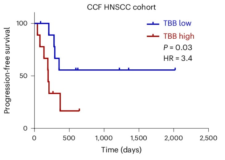
Further splitting patients in a tumour bacterial burden (TBB) high versus low group using the methodology above, stark differences in immunotherapy therapy appeared. The figure below shows the difference in progression-free survival depending on TBB status.